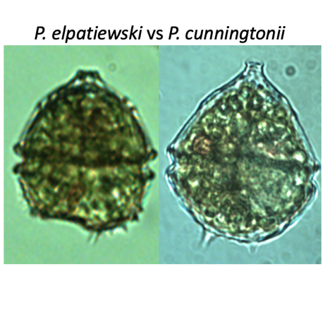
Morphological features: These two species are not so easy to tell apart: they are of the same size and occur in the water at the same time. Examining their thecal plate structure allows distinction between them: the plates differ in number and arrangement, especially when observed from above (Plate 1). However, without an SEM the plates are usually seen only on empty . To identify the living cells under the light microscope we examine (Plates 1, 2, 3):
(1) cell shape – P. elpatiewskyi is angular whereas P. cunningtonii is smooth and rounded, especially its quinquecuspidata forma;
(2) spines on – P. cunningtonii has 1-4 spines, more or less of the same size and evenly spaced. P. elpatiewskyi has 2-3 main spines and to the side of one of them a “brush” of smaller spines.
(3) hypotheca base - in P. elpatiewskyi it is flat to concave whereas that of P. cunningtonii is rounded.