- Phylum
- Cyanobacteriophyta
- Class
- Cyanophyceae
- Order
- Chroococcales
- Habitat
- planktonic, pelagic
- Distinctive features
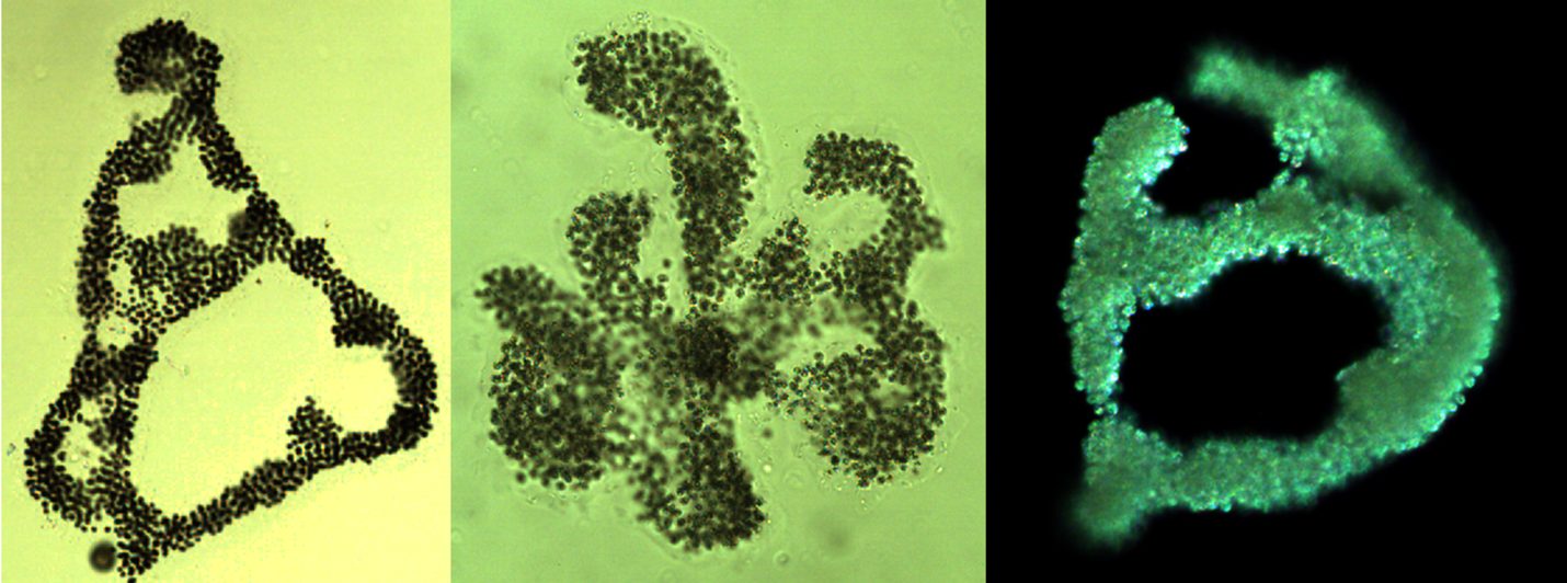
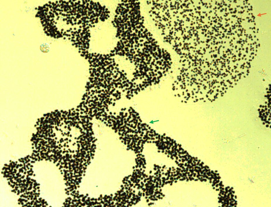
- irregular 3D colony shape often with holes. Spherical cells, similar size or slightly larger than those of M. flos-aquae. Usually, both species co-occur.
- Organization
- colonial
- Color
- green or brown
- Cell shape
- sphere
- Colony shape
- irregular
- Cell diameter (D)
- 2 – 5 μm, median: 3 μm (N=2500)
- Cell biovolume
- 4 – 65 μm3, median: 13 μm3
- Biovolume equation
- Sphere, V = 4/3 π (D/2)3
- Colony diameter
- 100-1000 μm
- Cells per colony
- 20-10000; median: 300
Morphological features
Colonies are net-like () with distinct holes or composed of sub-colonies with irregular outline, made of hundreds of irregularly gathered cells situated in a fine, colorless and diffuse mucilage with refractive outline. Cells are spherical, ~3 µm in diameter, with gas vesicles. Division in 3 perpendicular planes with a return development to the original spherical cell shape before the next succeeding division. Polymorphic species.
Ecology
The species was not counted prior to 1996, when Analyst 5 began counting, probably because earlier M. aeruginosa and M. flos-aquae were considered as two forma of a single species. Microcystis aeruginosa is less abundant than M. flos-aquae, but usually both species co-occur (Plate 2). The species is more abundant from March to June, less abundant from August to December (Fig. 2a). The number of cells per colony ranges 100-2000 with no distinct annual pattern (Fig. 2b). Cell volume is usually 10-40 µm3, being largest from May to August (Fig. 2c).
Physiological features
Like other species of Microcystis, M. aeruginosa produces microcystins, that are hepatotoxins, highly toxic to humans and mammals. It regulates its position in the water column by sinking and floating, using its gas vacuoles to float and carbohydrate ballast to sink. During calm weather it accumulates at the surface to form scums. When cultured in liquid medium, Microcystis colonies break to single cells.
Environmental conditions
M. aeruginosa biomass was indifferent to water temperature, pH, , chloride, calcium and concentrations (Fig. 3). Biomass was higher when water level was lower than -211 m a.s.l., Secchi was < 3.2 m, zeu < 10 m and nitrate < 0.4 mg L-1.
Cite this record as: Dr. Tamar Zohary, Dr. Alla Alster. 23 August 2026. Electronic publication. Israel Oceanographic & Limnological Research. https://kinneret-algae-atlas.org/ Searched on —.
Further reading
- Komárek J, Anagnostidis K. 1999. Cyanoprokaryota. 1. Chroococcales. In: Süßwasserflora von Mitteleuropa. Begründet von A. Pascher. Band 19/1. (Ettl H, Gärtner G, Heynig H, Mollenhauer D, Eds), pp. 1-548. Heidelberg & Berlin: Spektrum, Akademischer Verlag.
- McGregor G, Fabbro LD, Lobegeiger JS. 2007. Freshwater planktic Chroococcales (Cyanoprokaryota) from North-Eastern Australia: a morphological evaluation. Nova Hedwigia 84(3/4): 299-331.