- Phylum
- Cyanobacteriophyta
- Class
- Cyanophyceae
- Order
- Chroococcales
- Habitat
- planktonic, pelagic
- Distinctive features
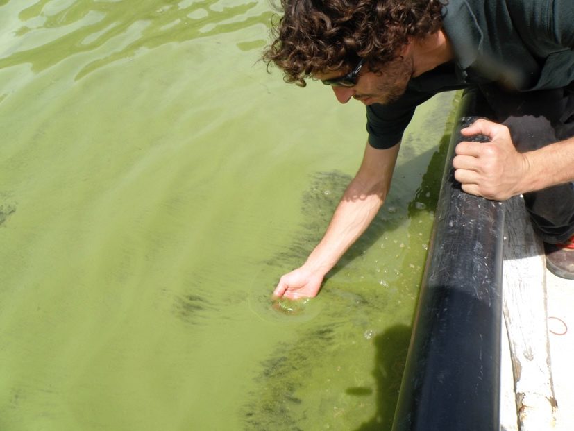
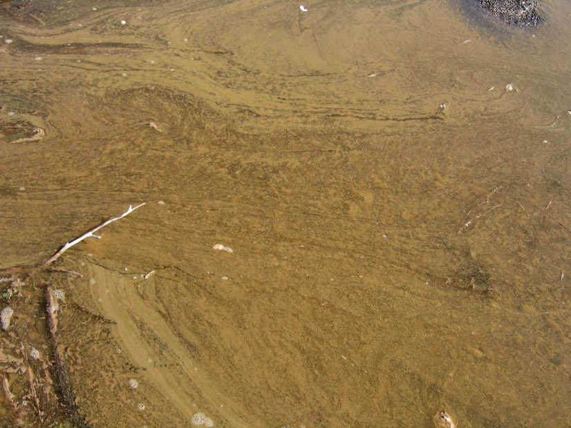
- abundant in Lake Kinneret in winter. During calm weather Microcystis colonies float to create a strongly pigmented (green or brown) surface film/scum, visible to the naked eye.
- Organization
- colonial
- Color
- green or brown
- Cell shape
- sphere
- Colony shape
- ranges from spherical to irregularly lobate with holes
Morphological features
In Lake Kinneret, several species of the genus Microcystis are important components of the winter plankton. They are colonial species made of small (3-7 µm) spherical cells (hemispherical after cell division) that contain gas vesicles, and are organized sparsely or densely in a common colorless mucilage, with no envelopes around individual cells. The colonies vary in shape from spherical to oval or elongate, lobate to irregular, in some species (with holes). Cell division is by binary fission in 3 perpendicular planes in regular cubic arrangement. Reproduction is by disintegration of colonies into small clusters of cells or even into single cells. The species occurring in Lake Kinneret are M. aeruginosa, M. botrys, M. flos-aquae, M. viridis and M. wesenbergii, each will be dealt separately. Here we address issues common to all those species.
Counting Microcystis cells is a difficult task, especially for M. flos-aquae and M. aeruginosa, colonies that are often made of hundreds to thousands of densely packed small-sized cells. For accurate cell counts it is essential to break the colonies into single cells (e.g. with a sonicator or high-speed blender) that can be dispersed evenly on a counting slide. But the procedure damages other more delicate species, and once broken into single cells it is impossible to count colonies and identify the cells to species level. We count Microcystis without breaking the colonies and take into consideration that the cell counts and therefore also biomass estimates for Microcystis species have a large measurement error.
Counting Microcystis cells is a difficult task, especially for M. flos-aquae and M. aeruginosa, colonies that are often made of hundreds to thousands of densely packed small-sized cells. For accurate cell counts it is essential to break the colonies into single cells (e.g. with a sonicator or high-speed blender) that can be dispersed evenly on a counting slide. But the procedure damages other more delicate species, and once broken into single cells it is impossible to count colonies and identify the cells to species level. We count Microcystis without breaking the colonies and take into consideration that the cell counts and therefore also biomass estimates for Microcystis species have a large measurement error.
Ecology
While in general “cyanobacteria blooms like it hot” (Paerl and Huisman 2008), and this is typical for Microcystis around the world, in Lake Kinneret Microcystis blooms occur in winter-spring, mostly between January and March. The main reason for this may be in Kinneret winter temperatures are warm enough, whereas in summer nutrient availability is low, and N-fixing cyanobacteria dominate. When abundant, on calm days, Microcystis floats to form green or brown surface films (Plates 1, 2). Those surface films are blown by gentle winds and accumulate near lee shores to create visible surface scums.
Microcystis blooms were reported to occur in Kinneret already in the 1960s, before the onset of the Kinneret monitoring program, with an exceptionally intensive bloom reported in winter 1964 (Pollingher and Kimor 1970; Serruya and Pollingher 1971, Pollingher 1978) and again in 1995 (Zohary 2004).
Microcystis blooms were reported to occur in Kinneret already in the 1960s, before the onset of the Kinneret monitoring program, with an exceptionally intensive bloom reported in winter 1964 (Pollingher and Kimor 1970; Serruya and Pollingher 1971, Pollingher 1978) and again in 1995 (Zohary 2004).
Physiological features
Microcystis contains gas vacuoles and can float and regulate its position in the water column. It floats to the surface where it dominates the light climate, suppressing competition from other species. When it has sufficient N, P and light it forms excessive biomasses that accumulate to form thick surface scums, in extreme cases - ‘hyperscums’ (Zohary and Breen 1989) that rot at the surface releasing pungent odors. But these occur in hypertrophic lakes, not in Kinneret.
Microcystis produces microcystins – hepato-toxins that are toxic to humans and other mammals. Hence, it is a health hazard, and its blooms impair water quality.
A studied characteristic of Microcystis in Lake Kinneret is its allelopathic interactions with Peridinium gatunense: when abundant, Microcystis inhibits the growth and photosynthesis of P. gatunense, and visa-versa – when the dinoflagellate is abundant, it inhibits Microcystis (Sukenik et al. 2002; Vardi et al. 2002).
When cultured in liquid medium, Microcystis colonies break to single cells.
Microcystis produces microcystins – hepato-toxins that are toxic to humans and other mammals. Hence, it is a health hazard, and its blooms impair water quality.
A studied characteristic of Microcystis in Lake Kinneret is its allelopathic interactions with Peridinium gatunense: when abundant, Microcystis inhibits the growth and photosynthesis of P. gatunense, and visa-versa – when the dinoflagellate is abundant, it inhibits Microcystis (Sukenik et al. 2002; Vardi et al. 2002).
When cultured in liquid medium, Microcystis colonies break to single cells.
Cite this record as: Dr. Tamar Zohary, Dr. Alla Alster. 23 August 2026. Electronic publication. Israel Oceanographic & Limnological Research. https://kinneret-algae-atlas.org/ Searched on —.
Further reading
- Hadas O, Kaplan A, Sukenik A. 2015. Long-term changes in cyanobacteria populations in Lake Kinneret (Sea of Galilee), Israel: an eco-physiological outlook. Life. 5(1):418-431.
- Hozumi A, Ostrovsky I, Sukenik A, Gildor H. 2020. Turbulence regulation of Microcystis surface scum formation and dispersion during a cyanobacteria bloom event. Inland Waters 10(1):51-70.
- Kaplan-Levy RN, Alster-Gloukhovski A, Benyamini Y, Zohary T. 2016. Lake Kinneret phytoplankton: integrating classical and molecular taxonomy. Hydrobiologia 764(1):283-302.
- Ninio S, Lupo A, Viner-Mozzini Y, Zohary T, Sukenik A. (2020). Microcystis bloom dynamics – multiannual shift in species dominance. Harmful Algae 92: 101710. DOI: 10.1016/j.hal.2019.101710.
- Paerl HW, Huisman J. 2008. Blooms like it hot. Science. 320(5872):57-58.
- Pollingher U, Kimor B. 1970. Seasonal and bathymetric changes in the composition of the phytoplankton populations of Lake Tiberias based on biomass estimations during the years 1964-1967. State of Israel, Ministry of Agriculture, Department of Fisheries, Sea Fisheries Research Station.
- Pollingher U. 1978. Annual pattern of algal succession. In: Serruya C. (Ed). Lake Kinneret, vol 32, Monographiae Biologicae. Junk. Hugue pp 243-246.
- Schweitzer‐Natan O, Ofek‐Lalzar M, Sher D, Sukenik A. 2023. The microbial community spatially varies during a Microcystis bloom event in Lake Kinneret. Freshwater Biology. 68(2):349-63.
- Schweitzer-Natan O, Ofek-Lalzar M, Sher D, Sukenik A. 2019. Particle-associated microbial community in a subtropical lake during thermal mixing and phytoplankton succession. Frontiers in Microbiology 10:2142.
- Serruya C, Pollingher U. 1971. An attempt at forecasting the Peridinium bloom in Lake Kinneret (Lake Tiberias). Internationale Vereinigung für Theoretische und Angewandte Limnologie: Mitteilungen. 19(1):277-91.
- Sukenik A, Eshkol R, Livne A, Hadas O, Rom M, Tchernov D, Vardi A, Kaplan A. 2002. Inhibition of growth and photosynthesis of the dinoflagellate Peridinium gatunense by Microcystis sp.(cyanobacteria): a novel allelopathic mechanism. Limnology and Oceanography. 47:1656-1663.
- Sukenik A, Hadas O, Kaplan A. 2014. Chap. 12. Cyanobacteria. In: Zohary T, Sukenik A, Berman T, Nishri A. [eds] Lake Kinneret: Ecology and Management, pp. 213-226. Springer, Heidelberg.
- Sukenik A, Kaplan A. 2021. Cyanobacterial harmful algal blooms in aquatic ecosystems: A comprehensive outlook on current and emerging mitigation and control approaches. Microorganisms. 9(7):1472.
- Sukenik A, Rosin C, Teltsch B, Banker R, Carmeli S. 1998. Toxins from cyanobacteria and their potential impact on water quality of Lake Kinneret, Israel. Israel journal of plant sciences. 46(2):109-15.
- Vardi A, Schatz D, Beeri K, Motro U, Sukenik A, Levine A, Kaplan A. 2002. Dinoflagellate-cyanobacterium communication may determine the composition of phytoplankton assemblage in a mesotrophic lake. Current biology. 12:1767-72.
- Weisthal Algor S, Sukenik A, Carmeli S. 2024. Sulfated Aeruginosins from Lake Kinneret: Microcystis Bloom, Isolation, Structure Elucidation, and Biological Activity. Marine Drugs. 22(9):389.
- Zohary T. 2004. Changes to the phytoplankton assemblage of Lake Kinneret after decades of a predictable, repetitive pattern. Freshwater Biology 49: 1355-1371.
- Zohary T., Breen CM. 1989. Environmental factors favoring the formation of Microcystis aeruginosa hyperscums in a hypertrophic lake. Hydrobiologia 178: 179192.