- Phylum
- Euglenophyta
- Class
- Euglenophyceae
- Order
- Euglenales
- Habitat
- plankton, pelagic
- Distinctive features
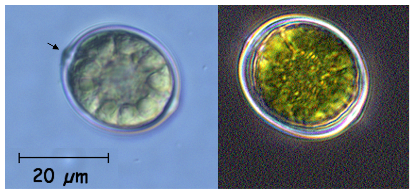
- Near-spherical flagellated cells encapsulated in a brown with a distinctive “mouth” opening through which the locomotive emerges.
- Organization
- single flagellated cells
- Color
- brown
- Cell shape
- sphaeroid
- Cell diameter (D)
- 8.9-15.5 μm (median=11.4 μm). Range reported is 5th-95th percentiles for 234 cells measured during 2004-2009.
- Cell biovolume
- 370 - 2000 μm3, median: 800 μm3.
- Biovolume equation
- sphere, V, μm3 = ¾ π (D/2)3
Morphological features
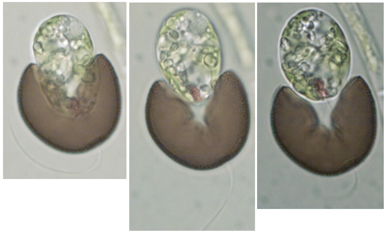
Trachelomonas cells are similar to those of Euglena, except that they are completely encased in a mucilaginous rigid envelope outside the plasma membrane called lorica (Plates 1, 2). This lorica is highly mineralized and dark brown in color. The cell is flagellated and capable of euglenoid movement within the lorica as well as swimming out of it (Plate 2). The lorica has a bottle-neck-shaped opening through which the flagellum emerges and through which the cell may swim out for cell division or when conditions require, then may swim back into it. Trachelomonas is distinguishable from other spherical unicellular species occurring in Lake Kinneret by the bottle-neck-shaped opening of its lorica, at the top part of the cell (arrow in Plate 1). This structure can be seen only when the cell is observed from the side, not when it is observed from above or below.
Ecology
Trachelomonas is the only euglenoid seen in Kinneret pelagic plankton samples quite often, although this genus too is never abundant, maximum cell density ever recorded was ~50/mL (Fig. 1). Trachelomonas can be viewed at any time of the year but tends to appear more often in spring (March-April) and rarely from August to October. (Fig. 2). Cell size varies only slightly, with no distinct annual pattern (Fig. 2).
Environmental conditions
The abundance of Trachelomonas spp. in the pelagic water of Lake Kinneret was indifferent to water temperature, ambient chloride concentration (Fig. 3), water level, pH, Ca, , Si, NO3, NH4 and Zmix/Zeu ratio (not shown). Higher abundances of the genus were recorded when depth-integrated (0-15 m) chlorophyll concentrations were < 500 mg m-2, and organic N > 0.15 mg N L-1 (Fig. 3).
Additional figures
Cite this record as: Dr. Tamar Zohary, Dr. Alla Alster. 16 June 2026. Electronic publication. Israel Oceanographic & Limnological Research. https://kinneret-algae-atlas.org/ Searched on —.