- Phylum
- Dinoflagellata
- Class
- Dinophyceae
- Order
- Thoracosphaerales
- Habitat
- plankton, littoral
- Distinctive features
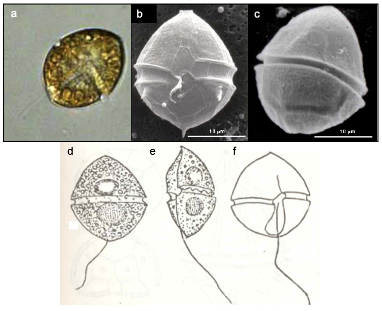
- convex-concave shape, seen when it circles around itself while swimming
- Organization
- flagellated single cells
- Color
- yellow to golden-brown
- Cell shape
- a prolate spheroid that was punched in its “belly” so that from view it is concave
- Cell diameter (D)
- 29-36 μm, median: 32 μm (N=32)
- Cell length (L)
- 32-42 μm, median: 38 μm (N=32)
- Cell biovolume
- 7,000-14,000 μm3, median: 10,000 μm3
- Biovolume equation
- V, μm3 = ((4/3)π L (D/2)2)/2 (half the volume of a prolate sphaeroid)
Morphological features
A medium-sized dinoflagellate. The flagellated cells are oval, with the side pressed in towards the other side as if it received a punch. As a result, the cell perimeter is convex when viewed from one side but concave when viewed from another (Plate 1). This concave/convex form is easily seen on a live, swimming cell: as it turns about its longitudinal axis its profile changes dramatically (Plate 1e). This feature is the most obvious for identifying this species. An additional diagnostic feature is the smooth and rounded cell shape (Plate 1). The is helmet-shaped and pointed and is approximately the same size as the broadly rounded . The chloroplasts are small and discoid or ribbon shaped. A () may be present. Resting cysts are spherical with their thick cell wall covered by dense hairlike projections (not shown).
Ecology
Naiadinium polonicum occurs in the Kinneret plankton but in low numbers, usually < 5 cells/mL-1 (Fig. 1). It is more abundant in shore samples than in pelagic waters. Its peak abundance is in April; it is absent in the water column during summer-fall (Fig. 2).
Environmental conditions
Higher abundance of N. polonicum is associated with the full range of , Ca, dissolved oxygen (not shown) and water temperatures, low concentrations of DIN (nitrate, ammonium) but intermediate concentrations of DON (Fig. 3). It tends to be more abundant at the lower salinities experienced in Lake Kinneret (Chloride < 250 mg/L) and at pH levels > 8.4 (Fig. 3).
Additional figures
Cite this record as: Dr. Tamar Zohary, Dr. Alla Alster. 16 June 2026. Electronic publication. Israel Oceanographic & Limnological Research. https://kinneret-algae-atlas.org/ Searched on —.
Further reading
- Hansen G, Flaim G. 2007. Dinoflagellates of the Trentino Province, Italy. J Limnol. 66:107-141.
- Penard E. 1891. Les Peridiniacees du Lac Leman. Bull. Trav. Soc. Bot. Geneve 6: 1-63.
- Pollingher U, Hickel B. 1991. Dinoflagellate associations in a subtropical lake (Lake Kinneret, Israel). Arch. Hydrobiol. 120: 267-285.
- Popovsky, J. & Pfiester, L.A. 1990. Süßwasserflora von Mitteleuropa. Dinophyceae (Dinoflagellida). Vol. 6 pp. 1-272. Jena & Stuttgart: Gustav Fischer.