- Phylum
- Chlorophyta
- Class
- Chlorophyceae
- Order
- Volvocales
- Habitat
- plankton
- Distinctive features
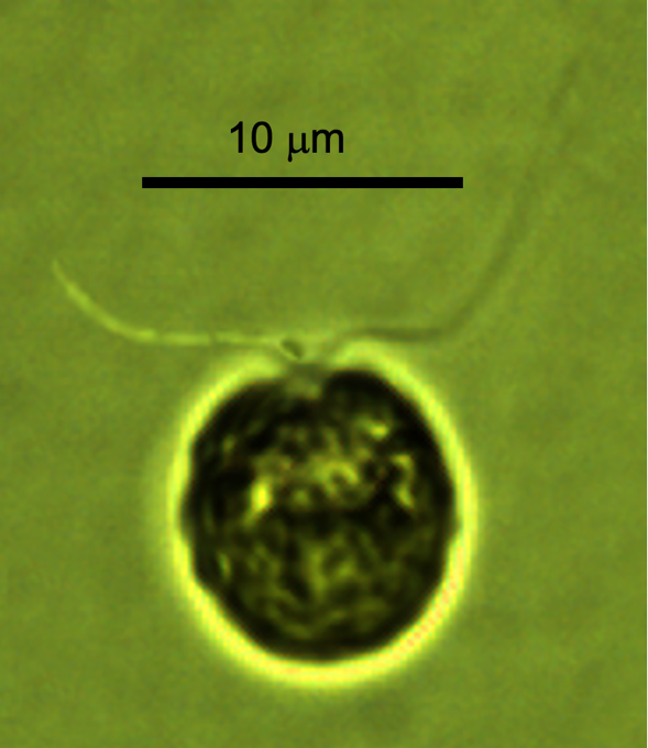
- small bi-flagellated single cells with cup-shaped chloroplast.
- Organization
- flagellated single cells
- Color
- green
- Cell shape
- spherical
- Cell diameter (D)
- 7 – 13 μm, median: 10 μm (N=562).
- Cell biovolume
- 200 – 1300 μm3, median: 520 μm3.
- Biovolume equation
- Sphere V, μm3 = 4/3 π (D/2)3.
Morphological features
Spherical flagellated green single cells, with two about same length as cell and a cup-shaped chloroplast. Several hundreds of species of Chlamydomonas were described, they are difficult to distinguish by light microscopy, we did not identify the Kinneret Chlamydomonas to species level, and it is possible that there is more than one species.
Ecology
Abundance data for this taxon was heavily biased by the person counting it: Technician 1 (1983 – 1995) counted more Chlamydomonas individuals in her samples than the two other technicians and Technician 2 counted the least (Fig. 1). This “technician effect”, dominant to other pattern in the cell abundance data, is a big limitation of long-term phytoplankton data sets based on microscope counts, it is not unique to Lake Kinneret. The discrepancies between technicians exist for all species, but at varying levels, depending on the taxon being counted: less conspicuous species that are easier to misidentify are more susceptible. For the annual cycle (Fig. 2) we present only data counted by Technician 3. It shows Chlamydomonas spp. are present in the water at all times of the year, with a tendency to higher abundance from October till February. Cell size remains close to constant over time, with no apparent annual pattern.
Environmental conditions
The Kinneret data for Chlamydomonas for 2013-2020 (Technician 3) do not show distinct correlations with any of the environmental factors we examined: it seems indifferent to water temperature, mixed layer depth, euphotic depth, Secchi depth, chloride, nutrients (not shown).
Additional figures
Cite this record as: Dr. Tamar Zohary, Dr. Alla Alster. 16 June 2026. Electronic publication. Israel Oceanographic & Limnological Research. https://kinneret-algae-atlas.org/ Searched on —.